Dysplastic nevi
Home • Dermatology • Skin Cancer, Pre-Cancerous Lesions & Treatment • Dysplastic neviAbout
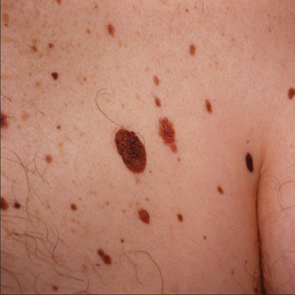
Dysplastic nevi, also known as atypical moles, are growths that have the potential to develop into melanoma. They are generally larger than ordinary moles and have irregular and indistinct borders. Their color can vary from one area to another with mixtures of tan, brown, red, and pink.
AboutSymptoms
Dysplastic nevi can be characterized by:
- Larger size than ordinary moles (generally larger than a pencil eraser)
- Irregular and poorly defined borders
- Mixed coloration, including shades of brown, black, red, or pink
- A flat or slightly raised appearance
Causes
While the exact cause of dysplastic nevi is not fully understood, they are believed to be influenced by genetic factors and sun exposure. Individuals with a family history of melanoma or who have multiple dysplastic nevi are at an increased risk of developing melanoma.
CausesTreatment Options
Management of dysplastic nevi, particularly when there’s a heightened risk of melanoma, requires a vigilant and strategic approach. Here’s a detailed look at the management strategies that can help in monitoring and addressing these atypical moles:
- Regular Skin Examinations – Individuals with dysplastic nevi should have their skin checked regularly by a dermatologist. These examinations typically occur every 6 to 12 months, depending on the number of moles and the presence of other risk factors for skin cancer.
- Photographic Monitoring – For patients with multiple dysplastic nevi, dermatologists might recommend total body photography. This involves taking detailed images of all body areas that have moles. These images serve as a baseline reference to track changes over time. This method is particularly useful for detecting new moles or changes in existing ones that might necessitate further examination or biopsy.
- Biopsy of Suspicious Moles – If a mole shows changes in size, color, shape, or symptom (such as itching or bleeding), a biopsy may be recommended. The procedure involves removing a part or all of the mole, which is then examined under a microscope by a pathologist to check for signs of cancer.
- Removal of Atypical Moles – In cases where a dysplastic nevus is found to be particularly suspicious or shares characteristics with melanoma, complete removal might be advised. This is done through surgical excision, where not only the mole but also some surrounding skin is removed to ensure all potentially dangerous cells are eliminated.
- Patient Education – Educating patients about the importance of self-examinations is a crucial part of managing dysplastic nevi. Patients learn how to perform regular self-examinations and are taught the “ABCDE” signs of melanoma (Asymmetry, Border, Color, Diameter, and Evolving), which can help them identify suspicious changes between professional skin exams.
- Sun Protection – Preventative measures against UV exposure are recommended to prevent new dysplastic nevi from forming and to limit changes in existing moles. This includes applying broad-spectrum sunscreen with an SPF of 30 or higher, wearing protective clothing, and avoiding peak sun exposure times.
- Advances – DermTech Pigmented Lesion Assay
By combining regular professional monitoring with vigilant self-care and protective measures, individuals with dysplastic nevi can effectively manage their condition and reduce their risk of developing melanoma.
Treatment Options